
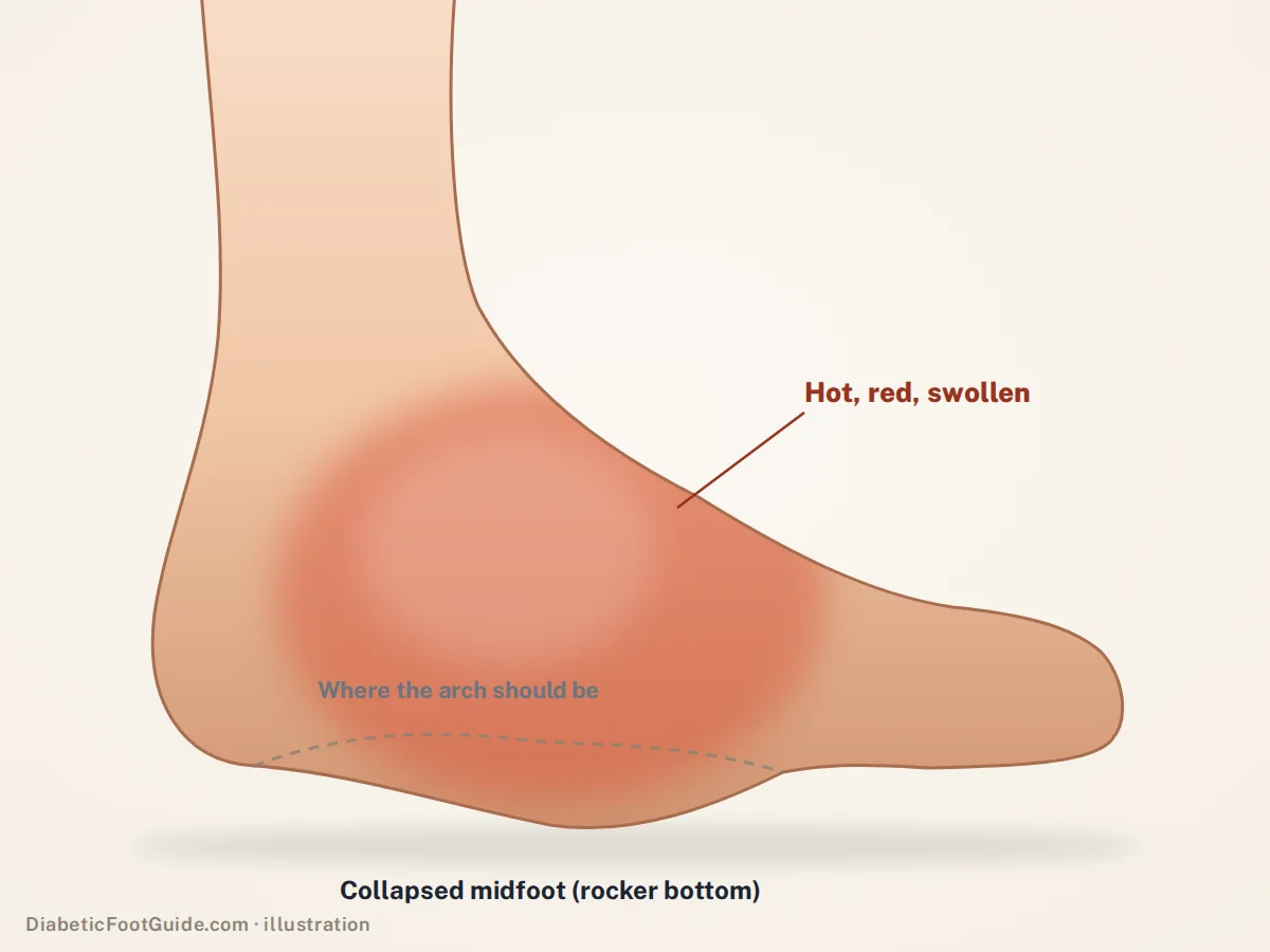
A hot, red, swollen foot in someone with diabetic nerve damage is a Charcot foot until proven otherwise. In one hospital series it was misdiagnosed in 63% of patients — as gout, as cellulitis, as a sprain — and the bones kept collapsing while everyone waited.
The short version
Charcot foot (Charcot neuroarthropathy, also called Charcot arthropathy or a Charcot joint) is a destructive breakdown of the bones and joints of the foot that happens in people whose protective sensation is gone — most often from diabetic neuropathy. Inflammation softens the bone, the person keeps walking on it because it does not hurt the way it should, and the arch collapses, sometimes within weeks.
The active phase is treated by taking every step off the foot, usually in a total contact cast, for months rather than weeks. Caught early, the foot can be preserved in a shape that still fits a shoe. Caught late, the treatment is reconstruction — or amputation.
The one thing to take from this page
If you have diabetes and neuropathy, and one foot becomes warm, red and swollen without an obvious wound, that foot needs to be seen within days, not at your next scheduled visit. It usually is not infected. It is often not a sprain. It very frequently is Charcot, and the window in which it can be stopped cheaply is measured in weeks.
The test you can do at home in ten seconds
Put the back of one hand on each foot, same spot, at the same time. A Charcot foot in its active phase is noticeably hotter than the other one — clinically the threshold used is a difference of more than about 2 °C between the same site on each foot, and in one study of the earliest stage that temperature difference was among the most significant findings, alongside a striking lack of pain.
One warm foot with no explanation is a reason to be examined. Two feet that feel the same are reassuring about this particular problem, and about nothing else.
Why it gets missed so often
Charcot is uncommon enough that most clinicians see it rarely, and it does an excellent impression of four things they see constantly. A survey of family and internal medicine physicians found that while most were aware of the condition, there was real disagreement about how to diagnose it and how to treat it without surgery.
| Mistaken for | What actually separates it |
|---|---|
| Cellulitis or infection | No wound, no portal of entry, and the person is not systemically unwell. Charcot redness typically fades when the leg is elevated for a few minutes; infection does not. |
| Gout | Gout is usually agonising and usually starts at the big toe joint. Charcot swelling centres on the midfoot or ankle, and hurts far less than a foot that shape should. |
| Deep vein thrombosis | DVT swells the calf as well as the foot and does not typically make one foot dramatically hotter than the other at the arch. |
| A sprain or a “bad step” | Many Charcot feet genuinely do start with a minor injury. The difference is that a sprain settles over a fortnight and this does not — it gets flatter. |
The trap underneath all four is the same. The foot does not hurt enough to be taken seriously, because the nerve that would have made it hurt is the reason this is happening in the first place.
What is physically going on
Three things happen at once. Neuropathy removes the pain that would normally stop you walking on an injured foot. An inflammatory cascade — often triggered by something small, a twist, a long day, a recent foot operation, an ulcer — floods the bone and switches on the cells that resorb it. And the bone itself, in long-standing diabetes, is already metabolically abnormal.
The result is bone that is temporarily soft, inside a foot that is still taking full body weight several thousand times a day, owned by someone who has no way of knowing. Joints dislocate. The midfoot drops. The arch inverts into the shape clinicians call a rocker-bottom foot, and the new bony prominence on the sole becomes a pressure point that ulcerates. That ulcer is how most Charcot feet eventually get infected, and infection in a deformed insensate foot is how they get amputated.
Charcot is not caused by walking on it, but walking on it is what makes it a deformity
The inflammation starts on its own. The collapse is mechanical. That is precisely why offloading — taking the weight off — is the treatment, and why it works even though it treats none of the underlying biology.
The stages
The classical description is the Eichenholtz classification, three stages based on what the X-ray shows. A fourth was added later at the front, because the most treatable version of this disease is the one where the X-ray is still normal.
-
Inflammation, normal X-ray
The foot is hot, swollen and red. Plain films show nothing wrong. MRI shows bone marrow oedema. This is the stage where the foot can be saved in a normal shape, and it is the stage that gets called gout and sent home. If you take one message from this section, take that one.
-
Fragmentation
The bone begins to break up and the joints subluxate. Now the X-ray is abnormal — debris, dislocation, loss of arch height. The foot is still hot. This is the destructive phase, and it is still an emergency for the foot.
-
Coalescence
Inflammation starts to settle. The temperature difference narrows. Bone fragments begin to knit and the foot starts to stabilise in whatever shape the fragmentation phase left it in.
-
Consolidation and remodelling
The foot is no longer hot. The bones have fused into a stable, permanent structure. If that structure is plantigrade — if it sits flat and takes weight sensibly — the outcome is footwear. If it is not, the outcome is reconstruction.
Stages are a description of a process, not a schedule. People move through them at very different speeds, and the transition is judged clinically by the foot cooling down and by serial imaging, not by the calendar.
How it is diagnosed
The examination first
Neuropathy confirmed with a 10-gram monofilament, a warm swollen foot, usually bounding pulses, and swelling that goes down overnight and comes back during the day.
Skin temperature
Infrared thermometry comparing identical sites on both feet. The same measurement is later used to decide when the cast can come off.
Plain X-rays
Essential, and normal in the earliest stage. A normal X-ray does not rule this out. That single sentence prevents a great deal of harm.
MRI
Detects the bone marrow oedema of stage 0 before anything appears on film. It is also what helps separate Charcot from osteomyelitis, which is the genuinely hard call.
Bloods are useful mostly for what they rule out. Inflammatory markers are often unremarkable in Charcot and raised in infection, but not reliably enough to decide on alone — particularly when both are present, which happens.
Treatment: offloading, and how long it really takes
There is no drug that reverses Charcot. Bisphosphonates and other bone-active agents have been trialled and none has become standard. The treatment is mechanical: get the load off the foot until the inflammation resolves, and hold the foot in a sensible shape while that happens.
The standard for doing that is the total contact cast — a close-fitting cast that spreads pressure across the whole lower leg and foot instead of concentrating it under the collapsing midfoot. It is changed regularly, both because limbs shrink as swelling settles and because a cast on an insensate leg has to be inspected for rubbing that the patient cannot feel.
Four months is the median, not the maximum, and it is the single fact patients are least prepared for. Being told “you will be in a cast until this foot stops being hot, and that may be most of a year” is hard to hear — but it is a great deal easier than what happens when the cast comes off early. Casting is also known to be underused; part of the reason is that it is skilled, slow work that clinics are not always set up to deliver.
Why a removable boot is not the same thing
A walking boot only offloads a foot while it is on. A total contact cast cannot be taken off to go to the bathroom at 2am, and that difference is most of its effect. Removable devices are used, but usually once the foot is cooling, or when a cast genuinely cannot be applied.
When surgery is on the table
Surgery is not the first move in an actively inflamed Charcot foot, and operating into that inflammation has its own risks. It becomes the right answer when the foot has consolidated into a shape that cannot safely take weight, when a bony prominence keeps ulcerating despite proper footwear, or when the ankle is unstable enough that bracing cannot control it.
The goal of reconstruction is not a normal-looking foot. It is a plantigrade one — a foot that sits flat, distributes load across an area rather than a point, and fits into a shoe without generating the next ulcer. That is achieved with fusions, osteotomies, removal of the offending prominence, and fixation that may be internal, external, or both. Recovery is long and the fixation frames are conspicuous, which is worth knowing before rather than after.
What happens after the foot has consolidated
A healed Charcot foot is a permanently higher-risk foot. It keeps the deformity, it keeps the neuropathy, and roughly one person in nine develops the same process on the other side. Lifelong management is mostly about footwear and inspection.
- Custom accommodative orthoses and therapeutic footwear. A rocker-bottom foot cannot be managed with an off-the-shelf insole; the device has to be built to accommodate the shape rather than correct it. Foot deformity is one of the qualifying conditions under the Medicare therapeutic shoe benefit, which is covered in detail on our diabetic shoes page — including the certification rule that catches most people out.
- A CROW walker — a custom clamshell brace — for feet that need more control than a shoe can give.
- Daily inspection of both feet, because the next problem will not announce itself with pain. If the skin over the prominence is red at the end of the day, the shoe is wrong.
- Prompt review of any new warmth or swelling, on either side.
Be seen the same day if any of this applies
- An open wound on a foot with a Charcot deformity, however small — see diabetic foot ulcers
- Fever, chills, or spreading redness up the leg
- Drainage, a bad smell, or a wound that probes down to something hard
- A sudden change in the shape of the foot, or a new inability to bear weight
- The other foot becoming warm and swollen
Common questions
Is Charcot foot the same as Charcot arthropathy or a Charcot joint?
Yes. Charcot foot, Charcot neuroarthropathy, Charcot arthropathy, neuropathic arthropathy and Charcot joint are all names for the same process: bones and joints breaking down in a foot that can no longer feel injury. The name comes from Jean-Martin Charcot, the French neurologist who described these joints in 1868 in patients with spinal cord damage from late syphilis. Today diabetes is by far the most common cause. The midfoot is the usual site, but the ankle and the back of the foot can be affected too, and a Charcot ankle is often harder to stabilize.
What triggers a Charcot foot?
Usually something unremarkable. A twisted ankle, a minor fracture, a recent foot operation, an existing ulcer, or no identifiable event at all. In one cohort, an ulcer or a traumatic injury preceded it in the large majority of cases. The trigger is not the disease — the disease is the inflammatory response to it in a foot that cannot feel and does not stop.
Does Charcot foot ever go away?
The active, inflamed phase resolves — that is what treatment achieves, and the foot stops being hot. The deformity left behind does not go away, and neither does the underlying neuropathy. So it is better thought of as a condition that burns out and leaves a permanently altered foot, rather than one that is cured.
What are the four stages of Charcot?
Stage 0 is inflammation with a normal X-ray. Stage 1 is fragmentation, where bone breaks up and joints dislocate. Stage 2 is coalescence, as the inflammation settles and fragments begin to knit. Stage 3 is consolidation, where the foot fuses into its final, permanent shape. Stage 0 was added to the original three-stage Eichenholtz description because it is the stage where treatment changes the outcome most.
Can a Charcot foot be fixed without surgery?
Frequently, yes — if it is caught early. Non-surgical treatment means prolonged offloading, most often in a total contact cast, followed by custom footwear or bracing for life. Surgery becomes necessary when the consolidated foot cannot take weight safely, keeps ulcerating over a bony prominence, or is too unstable to brace.
How long will I be in a cast?
Plan for months. A median of a little over four months has been reported for resolution of an acute Charcot foot, with a wide spread around that; coexisting osteoarthritis was associated with longer treatment. The cast comes off when the foot has cooled and the imaging supports it, not on a fixed date. What happens after the cast, from the CROW walker to custom shoes, is in our Charcot foot recovery guide.
Can I walk on a Charcot foot?
Not during the active phase, not without the offloading device, and not because it does not hurt. The absence of pain is the mechanism of the injury, not permission. What counts as safe weight-bearing during treatment is a decision for the clinician managing the cast, and it varies.
Sources
Clinical literature retrieved from PubMed.
- Hurst M, Shin L. Charcot neuroarthropathy: surgical and conservative treatment approaches. Semin Vasc Surg. 2025;38(1):74–84. doi:10.1053/j.semvascsurg.2025.01.004 — prevalence, 29% five-year mortality, and the gout / DVT / cellulitis misdiagnosis pattern.
- Griffiths DA, Kaminski MR. Duration of total contact casting for resolution of acute Charcot foot: a retrospective cohort study. J Foot Ankle Res. 2021;14(1):44. doi:10.1186/s13047-021-00477-5 — 63% misdiagnosis rate, two-month median delay, 4.3-month median casting duration, and the outcome split afterwards.
- Sebastian AP, Dasgupta R, Jebasingh F, et al. Clinical features, radiological characteristics and offloading modalities in stage 0 acute Charcot’s neuroarthropathy. Diabetes Metab Syndr. 2019;13(2):1081–1085. doi:10.1016/j.dsx.2019.01.008 — the greater-than-2 °C temperature difference and relative lack of pain in stage 0.
- Bilello J, Jupiter DC. A pilot survey: knowledge of Charcot neuroarthropathy among family and internal medicine practitioners. J Foot Ankle Surg. 2021;60(6):1204–1206. doi:10.1053/j.jfas.2020.11.011 — primary-care uncertainty about diagnosis and non-surgical treatment.
- Petrova NL, Edmonds ME. Conservative and pharmacologic treatments for the diabetic Charcot foot. Clin Podiatr Med Surg. 2016;34(1):15–24. doi:10.1016/j.cpm.2016.07.003 — total contact casting as the mainstay, its underuse, and the absence of an established drug therapy.
Related guides
Charcot foot recovery
How long the cast takes, what the CROW boot does, and the footwear that follows.
Diabetic foot ulcers
Why offloading is the treatment, and what a wound open at six weeks means.
Diabetic foot infection
The signs, and the probe-to-bone test that changes the plan.
Diabetic shoes and inserts
Foot deformity is a qualifying condition under the Medicare benefit.
The ICD-10 codes
E11.610 on its own, and why the M14.67- Charcot’s joint codes do not go with it.
Swollen feet and diabetes
Both feet usually points somewhere systemic. One warm, red foot is a different question and a much shorter clock.
Seen in Michigan for a hot, swollen foot
Balance Foot & Ankle sees Charcot, diabetic wounds and at-risk feet at two offices — Howell and Bloomfield Hills. If one foot is warm, red and swollen and you have neuropathy, that is a same-week problem.

Dr. Tom Biernacki is a double board-certified podiatric foot and ankle surgeon practising in Michigan. He treats diabetic wounds, Charcot neuroarthropathy and at-risk feet daily, and writes here to put the clinical reasoning behind that care in front of the people living with it.