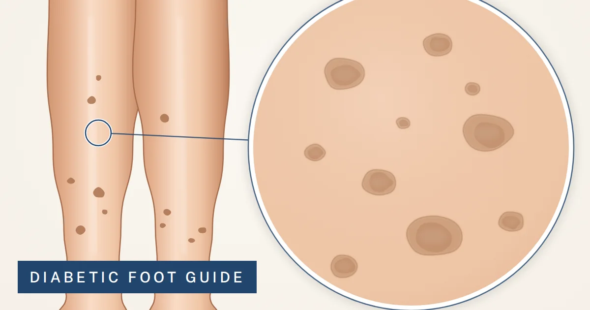
In someone with diabetes, small brown marks on the front of both shins are usually diabetic dermopathy, and they are harmless. The useful work is separating them from the marks that are not, and reading what the harmless ones say about your eyes, kidneys and nerves.
The short version
Diabetic dermopathy is not dangerous. The spots do not hurt or itch, need no treatment, and each fades on its own within a year or two while new ones appear.
What it signals is the reason to act. It is most common after years of diabetes and travels with eye, kidney and nerve damage, so treat it as a prompt to confirm those screenings are current.
Three look-alikes are not harmless: a red, hot, swollen, spreading leg (infection, same day); red or purple pinpoint spots that are raised or come with fever, bleeding gums or easy bruising (same day); and shiny yellow-brown shin plaques (necrobiosis lipoidica, which can ulcerate).
Spots on the legs: which one is this?
Position, texture and one leg versus both sort these faster than color.
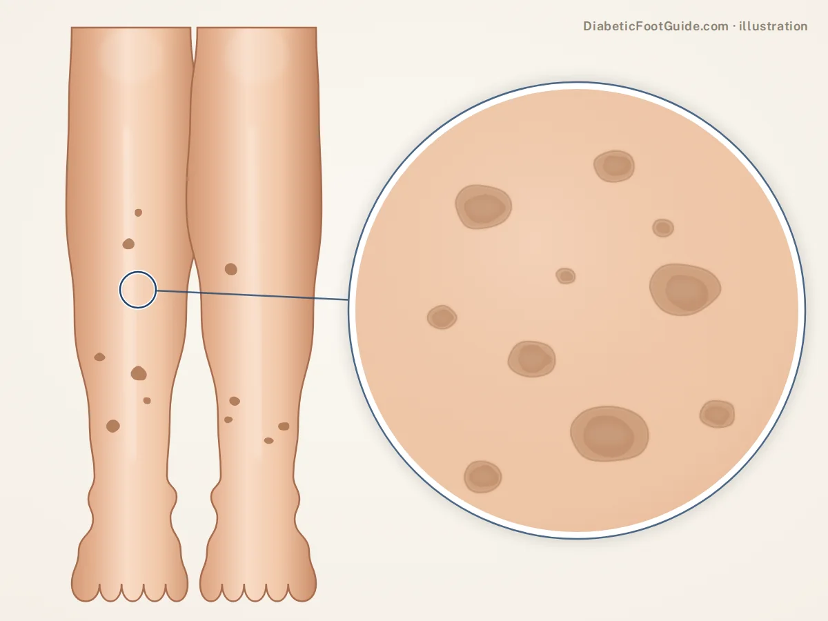
Diabetic dermopathy (shin spots)
Looks like: small, round, brown spots on the front of both shins, flat or slightly sunken.
Means: harmless scarring; a marker of long-standing diabetes and its complications.
Urgency: none for the skin; tell your diabetes care team.
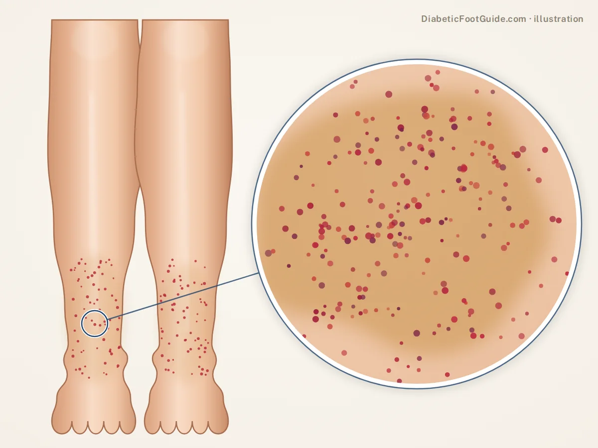
Red or purple pinpoint spots
Looks like: flat pinhead dots that stay visible when pressed, often on a golden-brown stain.
Means: usually capillaritis, a harmless leak from tiny vessels; less often low platelets or vasculitis.
Urgency: new, flat and feeling well: this week. Raised, or with fever, feeling unwell, bleeding gums or easy bruising: same day.
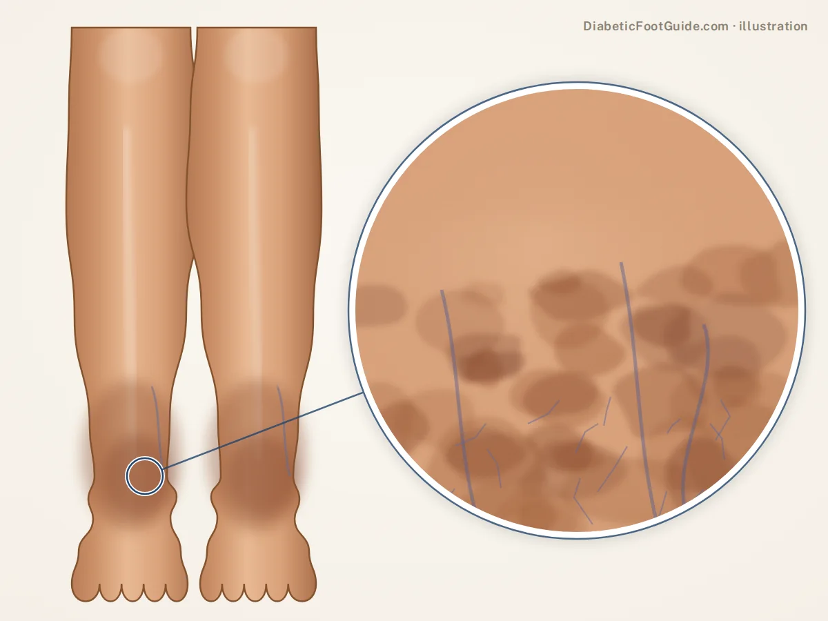
Rust-brown staining around the ankles
Looks like: a mottled brown band above the inner ankle bones, often with swelling and itch.
Means: iron from blood leaking out of overloaded veins. Venous disease, not diabetes.
Urgency: a vein assessment within weeks; this week if the skin weeps or opens.

Necrobiosis lipoidica
Looks like: larger, shiny, yellow-brown shin plaques with visible vessels and a red-purple rim.
Means: an uncommon diabetes-linked skin disease that can ulcerate.
Urgency: a dermatologist within weeks; this week if it has opened.
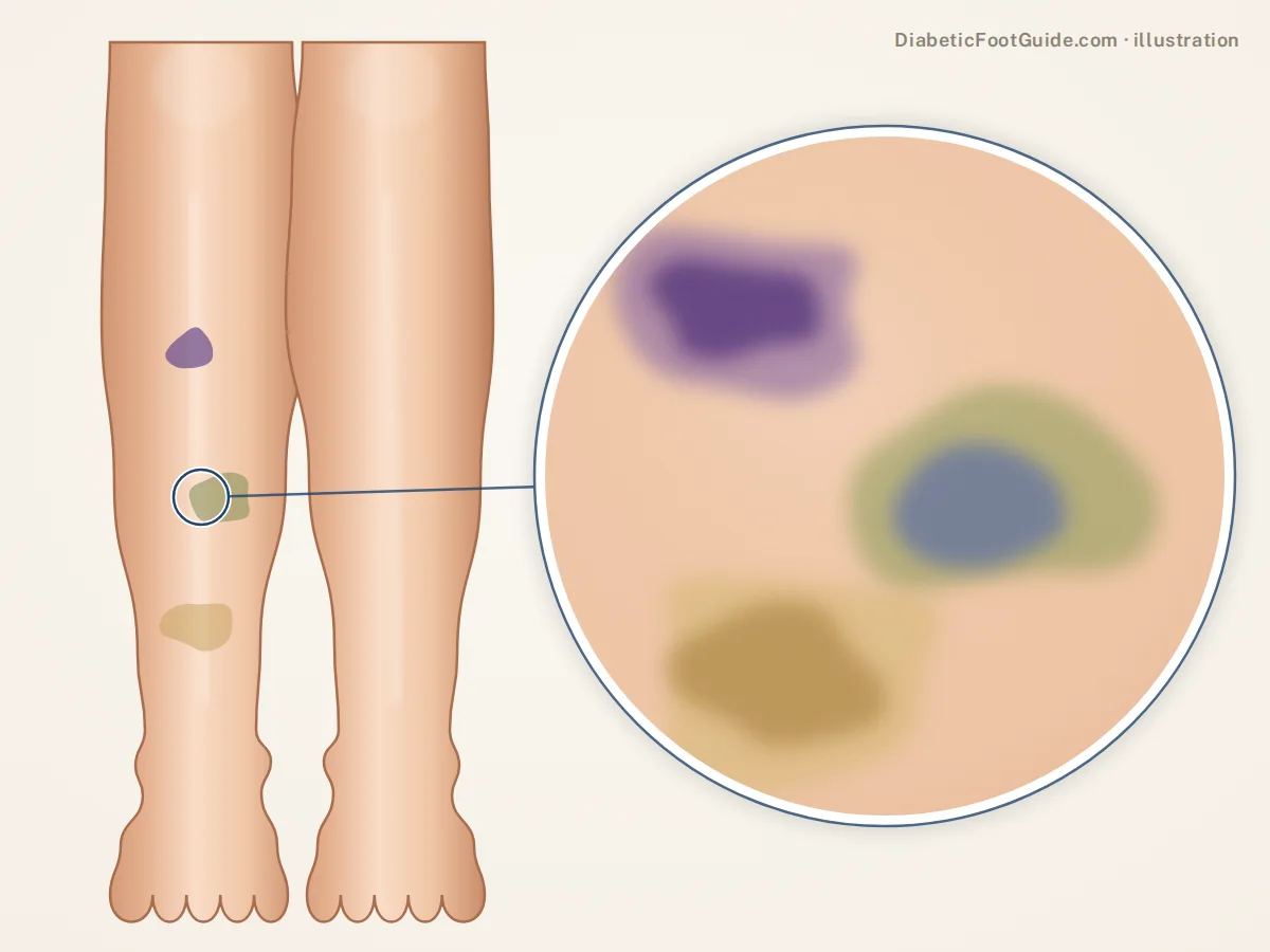
Bruises
Looks like: purple-blue patches that turn green, then yellow.
Means: a knock. Blood thinners enlarge bruises, and neuropathy means you may not have felt it.
Urgency: routine, but check for broken skin. Same day with nosebleeds, bleeding gums or pinpoint spots.
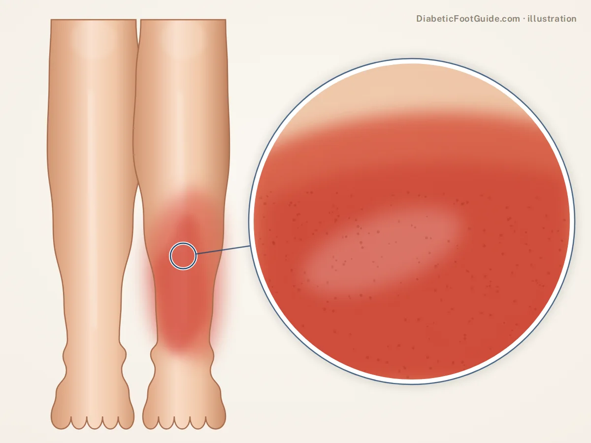
Cellulitis: red, hot, spreading
Looks like: one leg red (dusky purple on darker skin), swollen, warm and tender, spreading over hours.
Means: bacterial infection, often entering through a crack on the foot.
Urgency: same day. With fever, chills or red streaks, the emergency department.
Red spots on the legs with diabetes
Most red spots on a diabetic leg are not caused by diabetes. Three checks narrow them down.
- Press a clear glass on it. Inflamed skin fades under pressure; blood that has leaked into the skin does not. That sorts mechanism, not danger: cellulitis fades, harmless capillaritis does not.
- Flat or raised? Flat pinpoint dots on a golden-brown stain over the lower legs are usually capillaritis. It comes and goes and is harmless, though a first episode deserves a blood count to exclude low platelets. Reported triggers include aspirin and the diabetes drug glipizide. Purple bumps you can feel suggest vasculitis and need assessing the same day.
- One leg or both? Cellulitis usually takes one leg. Red, itchy, scaly skin around both ankles is more often stasis dermatitis from the veins, the condition most often mistaken for cellulitis. More than one in ten cellulitis diagnoses turn out to be wrong.
Two red patterns are tied to diabetes: a new dermopathy spot, red or purple before it browns, and eruptive xanthomas, sudden crops of yellow-topped bumps on a red base over the knees, elbows or buttocks. Xanthomas mean very high triglycerides and need fasting blood tests within days.
Waiting to be seen for a warm red patch? Outline it in pen and note the time; redness crossing the line means it is spreading.
On darker skin, do not wait for red
On brown and black skin, inflammation often looks purple, dark brown or gray, and infection gets missed while everyone waits for redness. Dermopathy spots may look dark brown to nearly black. Judge by warmth, swelling, tenderness and change, comparing both legs with the back of your hand.
Dark spots on the legs and ankle discoloration
Brown and dark marks sort mostly by position.
- Front of both shins, small and round: dermopathy.
- Around the inner ankles, spreading upward: venous staining.
- Golden-brown flecks with pinpoint dots: old capillaritis.
- Where you scratched, scraped or were bitten: post-inflammatory darkening, which fades over months, more slowly on darker skin.
- One new, changing or irregular dark spot: a skin-cancer check, not a diabetes question. The legs are a common site for melanoma, particularly in women.
For discoloration on the feet themselves, see brown spots on the feet.
Ankle discoloration with diabetes is usually the veins
When vein valves fail, pressure builds, red cells leak out of small vessels, and the iron they leave, hemosiderin, stains the skin rust-brown, first just above the inner ankle. Swelling, itch and scaly skin tend to come with it. Diabetes is not the cause; heart failure, kidney failure and obesity add to the risk.
The stain fades slowly, if at all. Compression is the mainstay of treatment, but on a diabetic leg the arteries should be checked first (see swollen feet and diabetes).
Two other ankle colors matter. A foot that turns deep red when hanging down and pale when raised points at the arteries. One red, warm, swollen ankle with no wound and no remembered injury, on a numb foot, is Charcot foot until proven otherwise and needs examining within days.
Diabetic dermopathy: how the spots start, change and fade
Often called the most common skin sign of diabetes, dermopathy runs the same course in every spot, so the shins show several stages at once.
| Phase | What you see | Timing |
|---|---|---|
| New | Round or oval, red to purple, sometimes scaly; does not fade when pressed | Appears without a remembered injury, often unnoticed |
| Established | Well-defined brown patch, usually under 1 cm, flat or slightly sunken; the deeper the dip, the darker | Months |
| Fading | The brown clears, leaving a pale, thin, slightly depressed mark like a small scar | Typically 12 to 24 months after the spot appeared |
Where and who: the front of the shins, on both legs but not in mirror image, in people who have had diabetes of either type for years, more often after 50. Spot numbers rise with diabetes duration.
Why brown, and why a scar: the color is iron from leaked red cells plus extra melanin. Blood flow inside a spot is higher than around it, the pattern of scar tissue, so the leading view is a small scar from poor healing in skin whose small vessels diabetes has affected.
What it travels with: among 173 patients, dermopathy was linked with each of neuropathy, nephropathy and retinopathy, and the more of those a person had, the likelier the spots. A coronary artery disease link has been reported too, and in some series the spots appeared before the eye and kidney changes.
What the spots should prompt
- A dilated eye exam, if the last one was over a year ago
- Kidney tests: urine albumin-to-creatinine ratio and eGFR
- A diabetic foot exam with monofilament and pulse checks
- Blood pressure, cholesterol and smoking, given the heart link
- Never diagnosed with diabetes? An A1C test
What helps, and what to leave alone
No cream, pill or procedure has been shown to clear dermopathy, and the spots do not need clearing. The aim is to protect the skin, limit the contrast and control the diabetes behind it.
- Guard the shins from car doors, dishwasher doors and bed frames, and clean and cover any scrape.
- Moisturize daily with a plain, fragrance-free lotion, which keeps the surrounding skin from cracking and itching. Shop fragrance-free body lotion on Amazon
- Use sunscreen or clothing. Sun deepens brown marks, and the pale skin of old spots does not tan evenly.
- Camouflage them if you like. Waterproof leg makeup hides them well. Shop waterproof leg and body makeup on Amazon
- Work on glucose control. Existing spots respond unpredictably, but control pays off in the eyes, kidneys and nerves.
Leave these on the shelf
- Scrubs, pumice and microdermabrasion. The skin in a spot is already thin, and a scraped diabetic shin heals slowly.
- Bleaching and steroid creams. No evidence of benefit, and steroids thin skin further.
- Products sold as a cure. None has meaningful evidence behind it.
Have it looked at if
- A spot is raised, lumpy, growing or colored differently from the rest
- Anything on the shin has opened, crusted or is weeping
- A patch is bigger than a coin, or has a waxy, yellowish surface
- The skin around it is warm, swollen or getting redder
How urgent is it?
Find the row that matches what you see.
- Seen the same day
One leg red or dusky, hot, swollen and spreading. Or red-purple spots that are raised, or come with fever, feeling unwell, bleeding gums or unexplained bruising.
Same-day care; with fever or chills, the emergency department. Diabetic foot infection →
- Seen this week
New flat pinpoint spots while you feel well. A spot or plaque that has opened. Shiny yellow-brown plaques. Weeping or hardening ankle skin. A new or changing dark spot.
Primary care, podiatry or dermatology. Say what changed and when.
- Mention at the next visit
Small, painless brown spots on the front of both shins, unchanged for months.
Use them as the cue to check when you last had eye, kidney and foot checks.
Other skin signs of diabetes worth knowing
- Acanthosis nigricans: velvety, darkened skin in the neck, armpit or groin folds, tied to high insulin and a raised chance of type 2 diabetes.
- Skin tags: soft flaps on narrow stalks in the same folds, also linked with high insulin.
- Diabetic blisters: large, clear, painless blisters on the lower legs or feet with no rub to explain them. See blisters on a diabetic foot.
- Fungal infections: between the toes, across the soles and in the nails. See rashes on diabetic feet.
Common questions
What does diabetic dermopathy look like?
Small round or oval patches on the front of the shins, usually under a centimeter, light to dark brown, flat or slightly sunken. New ones can start red or purple. There are usually several, on both legs, and they neither hurt nor itch.
Is diabetic dermopathy dangerous?
No. The spots cause no symptoms, need no treatment and fade on their own. What matters is what they accompany: long-standing diabetes, eye, kidney and nerve damage, and a reported link with coronary artery disease.
Does diabetic dermopathy go away?
Each spot does, usually within 12 to 24 months, often leaving a faint pale dent. New spots tend to appear as old ones fade, so the shins seldom look completely clear. No treatment reliably hurries the fading.
What causes red spots on the legs with diabetes?
Usually not diabetes itself. Capillaritis, stasis dermatitis, bruises and bites are common, and new dermopathy spots start reddish. Same-day care is for a hot, spreading red area on one leg, or red-purple spots that are raised or come with fever or bleeding.
Why are my ankles discolored?
Rust-brown staining around the inner ankles usually comes from the veins: red cells leak out under pressure and leave iron in the skin. It deserves a vein assessment. One red, warm, swollen ankle on a numb foot is a different problem, to be examined within days.
Is there a cream for diabetic dermopathy?
No cream has been shown to clear it. A plain moisturizer, sunscreen and leg makeup are the useful products. Skip bleaching and steroid creams.
Can shin spots be a sign of prediabetes or undiagnosed diabetes?
They can, which is why dermatology reviews advise testing for diabetes when classic shin spots appear; an A1C test settles it. They are not proof: Swedish population studies found similar marks in about 2% of people without diabetes. Acanthosis nigricans, velvety darkening in the neck or armpit folds, is the better-known early warning.
Sources
Clinical literature retrieved from PubMed.
- Morgan AJ, Schwartz RA. Diabetic dermopathy: A subtle sign with grave implications. J Am Acad Dermatol. 2008;58(3):447-451. doi:10.1016/j.jaad.2007.11.013 — links with microvascular complications and coronary disease, and testing for diabetes when dermopathy is found.
- Shemer A, Bergman R, Linn S, et al. Diabetic dermopathy and internal complications in diabetes mellitus. Int J Dermatol. 1998;37(2):113-115. doi:10.1046/j.1365-4362.1998.00273.x — age, diabetes duration and complication count in 173 patients.
- Naik PP, Farrukh SN. Clinical Significance of Diabetic Dermatopathy. Diabetes Metab Syndr Obes. 2020;13:4823-4827. doi:10.2147/DMSO.S286887 — the red-to-brown course, 12-to-24-month lifespan, iron and melanin pigment, scar-like blood flow, look-alikes, and no proven treatment beyond camouflage.
- Duff M, Demidova O, Blackburn S, et al. Cutaneous Manifestations of Diabetes Mellitus. Clin Diabetes. 2015;33(1):40-48. doi:10.2337/diaclin.33.1.40 — dermopathy spots under 1 cm that clear in one to two years, their frequency without diabetes, and the other skin signs described here.
- Reid SD, Ladizinski B, Lee K, et al. Update on necrobiosis lipoidica: A review of etiology, diagnosis, and treatment options. J Am Acad Dermatol. 2013;69(5):783-791. doi:10.1016/j.jaad.2013.05.034 — necrobiosis lipoidica plaques and their frequent ulceration.
- Spigariolo CB, Giacalone S, Nazzaro G. Pigmented Purpuric Dermatoses: A Complete Narrative Review. J Clin Med. 2021;10(11):2283. doi:10.3390/jcm10112283 — capillaritis, its drug triggers, the platelet check, and palpable spots as vasculitis.
- Yosipovitch G, Nedorost ST, Silverberg JI, et al. Stasis Dermatitis: An Overview of Its Clinical Presentation, Pathogenesis, and Management. Am J Clin Dermatol. 2023;24(2):275-286. doi:10.1007/s40257-022-00753-5 — hemosiderin staining at the inner ankle, its risk factors, compression, and misdiagnosis as cellulitis.
Related guides
Brown spots on the feet
Marks on the feet and under the nails.
Rashes on diabetic feet
Fungal, dry-skin and circulation changes below the ankle.
Swollen feet and diabetes
Venous swelling, and why compression waits for a circulation check.
The diabetic foot exam
The check to book once dermopathy appears.
Diabetic feet pictures
What diabetic foot changes look like.
Skin changes on your legs or feet, in Michigan?
Balance Foot & Ankle assesses diabetic skin changes, circulation problems and wounds in Howell and Bloomfield Hills.

Dr. Tom Biernacki is a double board-certified podiatric foot and ankle surgeon practising in Michigan. He treats diabetic wounds, Charcot neuroarthropathy and at-risk feet daily, and writes here to put the clinical reasoning behind that care in front of the people living with it.