Not every ulcer on a foot is diabetic. Where it sits, what the edge looks like and whether it hurts will usually tell you which of four kinds it is — and the four have opposite treatments, which is why getting this right matters more than the dressing does.
The short version
Four families cover most foot and ankle ulcers: neuropathic (pressure on a numb foot), venous (blood struggling to leave the leg), arterial (blood struggling to arrive), and pressure (sustained load on immobile tissue, classically the heel).
The single most consequential distinction is venous versus arterial, because compression helps the first and can harm the second. Anyone putting a compression wrap on a leg without knowing which one they are looking at is guessing.
Telling them apart
| Neuropathic | Venous | Arterial | Pressure | |
|---|---|---|---|---|
| Where | Under pressure points — ball of the foot, tips or tops of toes, under a bony prominence | Above the inner ankle, the “gaiter” area | Toes, tips of toes, outer ankle, or anywhere with the least blood supply | Back of the heel, or over any bone in someone immobile |
| Shape and edge | Round, punched out, thick callus ringing it | Shallow, irregular, wet, with a ruddy base | Sharply demarcated, dry, pale or black base | Follows the shape of the pressure, often deeper than it looks |
| Pain | Often none | Aching, better with the leg raised | Often severe, worse when the leg is raised and better hanging down | Depends on sensation |
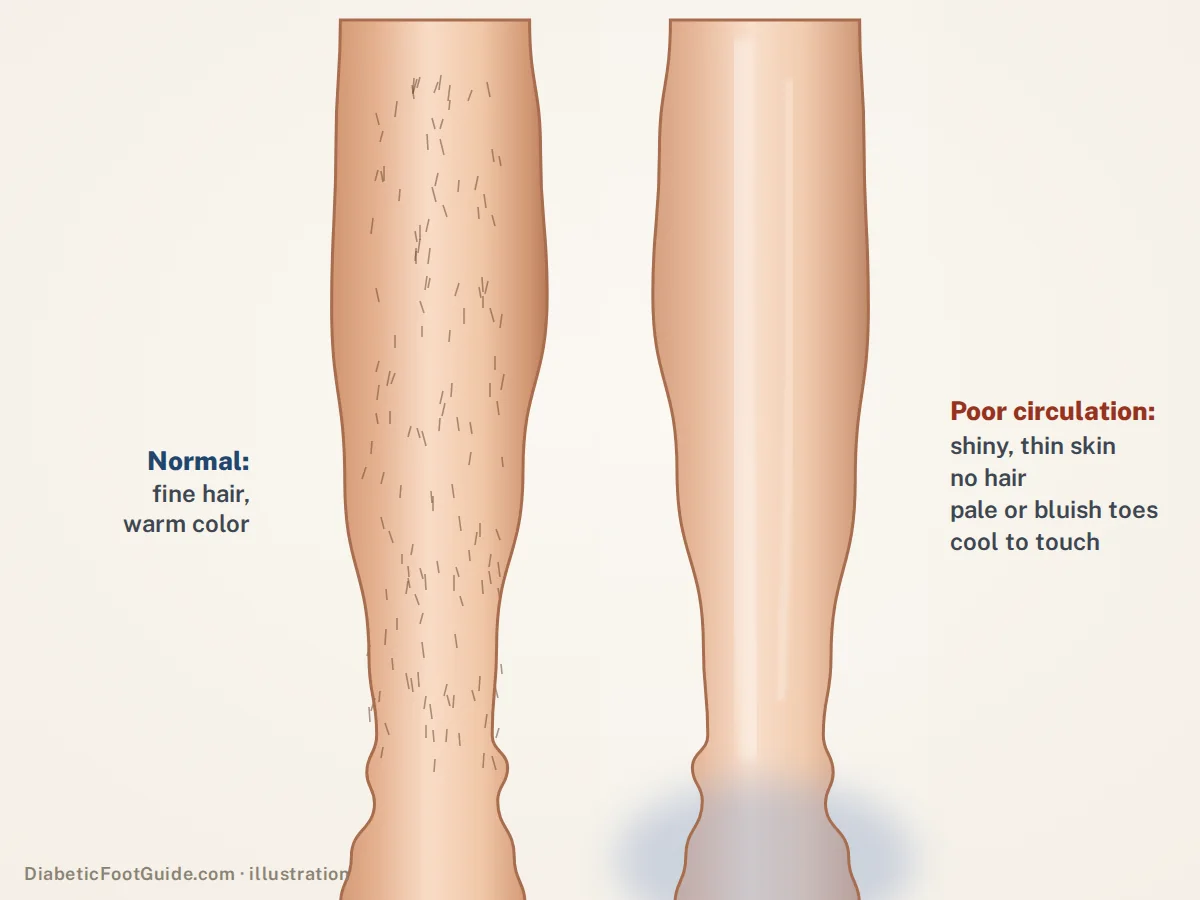
| Surrounding skin | Callus, dry skin, deformity | Brown staining, swelling, eczema, hardened tissue | Cool, shiny, hairless, thin, weak or absent pulses | Redness or bruising over the bone |
| Treatment centres on | Taking the pressure off | Compression, once arterial supply is confirmed | Restoring blood flow | Repositioning and offloading the heel |
The mistake that does real harm
Compression on a leg with significant arterial disease can reduce an already marginal blood supply. Compression is the treatment for venous ulceration and it should not go on until circulation has been assessed — and in a diabetic leg, assessed with toe pressures rather than an ankle-brachial index alone, because calcified vessels read falsely high.
Mixed ulcers are common, and that is the hard part
Real feet do not read the table. A great many ulcers are neuro-ischaemic — a pressure wound on a numb foot that also has poor inflow — and mixed venous-arterial disease is routine in older patients. The population presenting with foot ulcers has shifted over the last two decades away from purely neuropathic wounds towards mixed ones.
The practical consequence is that the first assessment is not “what does the wound look like” but “how much blood is getting here, and is anything still standing on it”. Everything else follows from those two answers.
The less common causes worth knowing exist
- Pyoderma gangrenosum — a rapidly enlarging, painful ulcer with a violet undermined edge, often in someone with inflammatory bowel disease or arthritis. Debriding it makes it worse, which is exactly the opposite of every other ulcer here.
- Vasculitis — multiple small, punched-out, painful ulcers, often with a rash, and usually a systemically unwell patient.
- Malignancy — any ulcer that has not healed despite correct treatment, or that has a rolled, everted edge or exuberant tissue, should be biopsied. A long-standing wound can transform.
- Infection as the primary cause — rather than a complication — including atypical organisms in the right exposure history.
- Sickle cell disease — classically ulcers around the ankles in a younger patient.
The rule that catches all of these
An ulcer that has not measurably improved in four to six weeks of correct treatment needs the diagnosis questioned, not the dressing changed. That is the point to reassess circulation, image for bone involvement, and consider biopsy.
What the assessment should include
Circulation
Pulses, ankle-brachial index, and toe pressures where the vessels may be calcified. This decides whether healing is possible at all.
Sensation
A 10-gram monofilament. Loss of protective sensation reframes the whole wound — and the patient’s ability to protect it.
The wound itself
Site, size, depth, edge, base, exudate, odour, and whether a probe reaches bone.
The leg above it
Swelling, staining, varicosities, skin changes — the venous story is written above the ankle, not in the wound.
Common questions
Can you get a foot ulcer without diabetes?
Yes, and it is common. Venous disease, arterial disease and pressure account for a large share of leg and foot ulcers in people who have never had diabetes. Neuropathy also has causes other than diabetes, so even a classically neuropathic-looking ulcer does not require a diabetes diagnosis.
How do I know if my ulcer is venous or arterial?
Position and pain are the strongest clues: venous ulcers sit above the inner ankle, ache, and feel better with the leg raised; arterial ulcers sit on the toes or outer ankle, often hurt badly, and feel better hanging down. But the distinction should be confirmed with pressure measurements, not decided on appearance, because the treatments conflict.
Why has my ulcer not healed after months of dressings?
Usually one of four things: the blood supply has never been properly assessed, the pressure has never actually come off it, there is infection in the bone, or the diagnosis is wrong. Dressings support healing; they do not cause it. Months without progress is a signal to re-open the diagnosis.
Should I put compression on it?
Not until someone has established that the arterial supply can tolerate it. Compression is the mainstay for venous ulceration and it is the wrong answer — and potentially a harmful one — in significant arterial disease. This is a decision for someone who has measured, not for a shelf in a pharmacy.
When should an ulcer be biopsied?
When it has failed to improve despite correct treatment, when the edge is rolled or heaped rather than sloping, when the tissue looks exuberant or friable, or when the story does not fit any of the common causes. A long-standing wound can become malignant, and the only way to know is to sample it.
Sources
Clinical literature retrieved from PubMed.
- Abbade LPF, Frade MAC, Pegas JRP, et al. Consensus on the diagnosis and management of chronic leg ulcers – Brazilian Society of Dermatology. Anais Brasileiros de Dermatologia. 2020;95:1-18. doi:10.1016/j.abd.2020.06.002 — venous, arterial and neuropathic causes accounting for about 90% of leg ulcers, the ABI thresholds, and compression contraindicated in severe arterial disease.
- O’Meara S, Cullum N, Nelson EA, et al. Compression for venous leg ulcers. Cochrane Database of Systematic Reviews. 2012;(11):CD000265. doi:10.1002/14651858.CD000265.pub3 — compression as the established treatment for venous leg ulceration.
- O’Donnell TF, Passman MA, Marston WA, et al. Management of venous leg ulcers: Clinical practice guidelines of the Society for Vascular Surgery and the American Venous Forum. Journal of Vascular Surgery. 2014;60(2):3S-59S. doi:10.1016/j.jvs.2014.04.049 — guideline management of venous leg ulcers, including arterial assessment before compression.
- Mervis JS, Phillips TJ. Pressure ulcers: Pathophysiology, epidemiology, risk factors, and presentation. Journal of the American Academy of Dermatology. 2019;81(4):881-890. doi:10.1016/j.jaad.2018.12.069 — pressure ulceration as a distinct mechanism from vascular and neuropathic ulcers.
- Armstrong DG, Boulton AJ, Bus SA. Diabetic Foot Ulcers and Their Recurrence. N Engl J Med. 2017;376(24):2367-2375. doi:10.1056/NEJMra1615439 — the neuropathic ulcer mechanism and its separation from ischaemic ulceration.
Related guides
Diabetic leg ulcers
Venous, arterial or neuropathic — where each sits on the leg, and why compression comes after the circulation check.
Diabetic foot ulcers
The neuropathic version, in detail.
Diabetic foot infection
What changes when any ulcer becomes infected.
The diabetic foot exam
Sensation and circulation testing, which decides the category.
Gangrene in the diabetic foot
Where an arterial ulcer ends up if inflow is not restored.
Wound care in Michigan
Balance Foot & Ankle assesses and treats foot and ankle ulcers of all four kinds in Howell and Bloomfield Hills, including circulation testing and offloading.

Dr. Tom Biernacki is a double board-certified podiatric foot and ankle surgeon practising in Michigan. He treats diabetic wounds, Charcot neuroarthropathy and at-risk feet daily, and writes here to put the clinical reasoning behind that care in front of the people living with it.