
A diabetic foot infection is diagnosed by looking at the foot, not by swabbing it. Every open wound grows bacteria; that is colonisation. Infection is what the tissue around the wound is doing — and in a diabetic foot it does it quietly.
The short version
Infection is a clinical diagnosis: two or more of local swelling, redness extending beyond the wound edge, tenderness, warmth, or purulent discharge. A culture tells you what is growing, not whether you are infected.
The question that changes the whole plan is whether it has reached bone. A probe that passes through the ulcer onto hard, gritty bone substantially raises the odds of osteomyelitis — and a normal early X-ray does not rule it out, because bone has to lose a great deal of mineral before it shows on film.
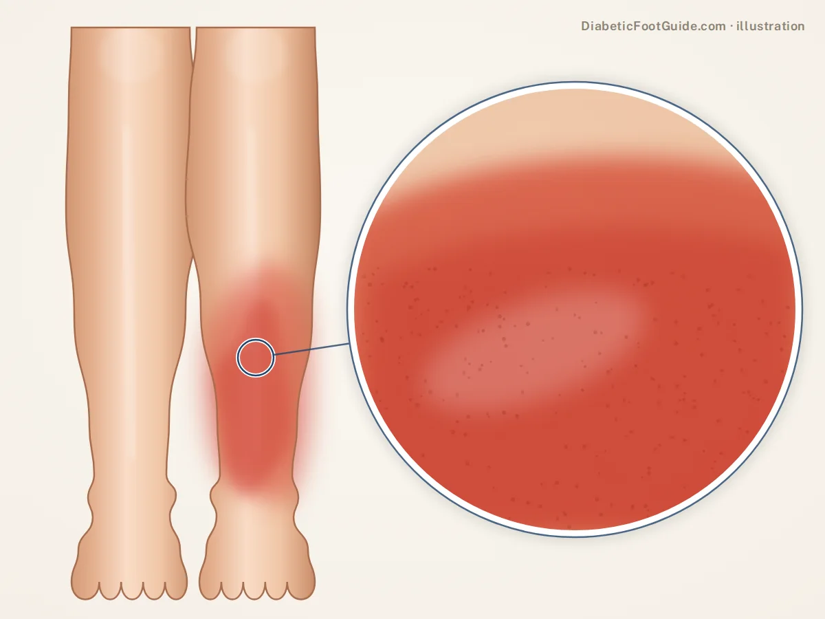
Why the usual signs are muted here
High glucose impairs how white cells find, engulf and kill bacteria. Neuropathy removes the pain that normally makes an infected foot impossible to walk on. Poor arterial supply limits both the immune cells and the antibiotics reaching the tissue. So a diabetic foot infection can be well advanced while the foot looks only moderately unhappy and the person feels systemically fine.
What the absence of fever actually tells you
Very little. Many people with a serious diabetic foot infection are afebrile with a normal white cell count. Judging severity on temperature alone under-calls this diagnosis routinely.
One signal worth knowing: blood sugars that suddenly become hard to control for no obvious reason often precede the visible infection. If your numbers have gone strange and there is a wound on your foot, those two facts belong in the same sentence.
How severity is judged
| Picture | What it usually means | Typical response |
|---|---|---|
| Wound present, no signs of inflammation | Not infected — colonised | Wound care and offloading. Antibiotics do not help and select resistance. |
| Redness extending a short distance around the wound, local warmth, swelling, tenderness or discharge | Mild infection, skin and subcutaneous tissue only | Usually oral antibiotics with close follow-up, plus debridement and offloading. |
| Redness extending further, or involvement deeper than skin — abscess, tendon, joint or bone | Moderate infection | Imaging, often surgical drainage or debridement, and a longer antibiotic course. |
| Any of the above plus fever, chills, confusion, vomiting, marked rise in blood sugars or metabolic instability | Severe — systemic | Hospital. This is an emergency. |
Two features move a foot up the scale regardless of how the skin looks: bone involvement, and poor arterial supply. An infected foot with inadequate circulation is a limb problem, not a skin problem.
The bone question
Probe to bone
A blunt sterile probe passed into the ulcer that meets hard, gritty resistance markedly raises the probability of osteomyelitis. It is a bedside test that costs nothing and is skipped constantly.
Plain X-ray
Always worth taking, and frequently normal early. Visible bone destruction means the process has been running for weeks. A normal film does not exclude it.
MRI
The imaging that settles it, showing marrow oedema before the bone erodes — and helping separate infection from a Charcot process, which is the genuinely difficult call.
Bone biopsy
The definitive answer, and the only way to know which organism is in the bone rather than which is on the surface. Surface swabs frequently disagree with bone cultures.
Why it matters: soft-tissue infection is usually days to a couple of weeks of antibiotics. Bone infection is weeks of targeted therapy, often surgery, and a materially different conversation about the foot’s future.
What good treatment looks like
- Drainage and debridement first where there is pus or dead tissue. Antibiotics do not sterilise an undrained abscess or dissolve necrotic tissue.
- Cultures taken from deep tissue, not from a surface swab. What grows on the surface of a chronic wound is often not what is causing the infection.
- Antibiotics chosen for severity and then narrowed once cultures return. Broad cover indefinitely is not a plan.
- Circulation assessed — because if blood is not reaching the tissue, neither is the antibiotic. Toe pressures rather than an ankle-brachial index alone, since calcified vessels read falsely high.
- Offloading continued throughout. The infection is being treated; the wound still will not close while it is being walked on. See diabetic foot ulcers.
- Glucose control tightened, because hyperglycaemia impairs the response you are relying on.
Emergency department today, not an appointment
- Fever, chills, feeling systemically unwell, confusion or vomiting
- Redness climbing the foot or up the leg
- Pus, a foul smell, or a wound that has suddenly enlarged
- Black tissue at the wound edge, or a toe turning dusky, cold or blue
- Crackling under the skin, or rapidly increasing swelling
- Blood sugars that have become uncontrollable with a wound on the foot
Common questions
How do I know if my diabetic foot wound is infected?
Look at the tissue around the wound rather than the wound itself: redness spreading beyond the edge, warmth, swelling, tenderness, or pus. Two or more of those signs is the working definition. A wound that simply looks unpleasant, or grows bacteria on a swab, is not automatically infected.
Can a diabetic foot infection be treated with antibiotics alone?
Mild, superficial infections often are — alongside debridement and offloading. Once there is an abscess, dead tissue, or bone involvement, antibiotics alone will not clear it. And if arterial supply is poor, the drug cannot reach the tissue in useful concentrations however good it is on paper.
What is the ICD-10 code for a diabetic foot infection?
There is no single code. You code the diabetes complication, the ulcer if one is present, and the specific infection — cellulitis or osteomyelitis with its own site and laterality. The structure is set out on our diabetic foot ICD-10 reference.
How serious is osteomyelitis in the foot?
Serious, and treatable. It generally means weeks of targeted antibiotics, often surgery to remove infected bone, and a real conversation about whether the foot can be reconstructed into a shape that takes weight. It is not automatically an amputation, and it is not something to manage with another course of oral antibiotics and hope.
Why does my doctor not want to swab the wound?
Because a surface swab of a chronic wound reports the bacteria living on it, which is nearly always a mixed population, and treating that list is not the same as treating an infection. When cultures are needed they are taken from deep tissue or bone, where the answer actually changes the antibiotic.
Sources
Clinical literature retrieved from PubMed.
- Senneville É, Albalawi Z, van Asten SA, et al. IWGDF/IDSA guidelines on the diagnosis and treatment of diabetes-related foot infections (IWGDF/IDSA 2023). Diabetes Metab Res Rev. 2024;40(3):e3687. doi:10.1002/dmrr.3687 — the IWGDF/IDSA severity classification and the treatment setting each grade implies.
- Schaper NC, van Netten JJ, Apelqvist J, et al. Practical guidelines on the prevention and management of diabetes-related foot disease (IWGDF 2023 update). Diabetes Metab Res Rev. 2024;40(3):e3657. doi:10.1002/dmrr.3657 — infection diagnosed on clinical signs rather than swab culture, and systemic signs being frequently absent.
- Lam K, van Asten SAV, Nguyen T, et al. Diagnostic Accuracy of Probe to Bone to Detect Osteomyelitis in the Diabetic Foot: A Systematic Review. Clin Infect Dis. 2016;63(7):944-948. doi:10.1093/cid/ciw445 — the diagnostic accuracy of probe-to-bone, pooled sensitivity 0.87 and specificity 0.83.
- Nawaz A, Torigian DA, Siegelman ES, et al. Diagnostic Performance of FDG-PET, MRI, and Plain Film Radiography (PFR) for the Diagnosis of Osteomyelitis in the Diabetic Foot. Mol Imaging Biol. 2009;12(3):335-342. doi:10.1007/s11307-009-0268-2 — plain radiography lacking the sensitivity to exclude osteomyelitis.
Related guides
Healing a diabetic wound faster
What speeds healing once the infection is under control.
Toe amputation with diabetes
Why it is done, how healing and walking go afterward, and what lowers the risk of another.
Diabetic foot ulcers
The wound the infection usually started in.
Gangrene in the diabetic foot
When infected tissue has already died.
Charcot foot
The hot swollen foot that is not infected, and how to tell.
The ICD-10 codes
How infection, ulcer and diabetes get coded together.
Swollen feet and diabetes
Both feet usually points somewhere systemic. One warm, red foot is a different question and a much shorter clock.
Foot infection care in Michigan
Balance Foot & Ankle manages diabetic foot infections, debridement and bone infection work-up in Howell and Bloomfield Hills. Say it is an infection when you book — those are seen first.

Dr. Tom Biernacki is a double board-certified podiatric foot and ankle surgeon practising in Michigan. He treats diabetic wounds, Charcot neuroarthropathy and at-risk feet daily, and writes here to put the clinical reasoning behind that care in front of the people living with it.