The ulcer people mean by “diabetic” forms on the foot, where a numb sole takes pressure it cannot feel. An ulcer on the leg — shin, calf or above the ankle — is usually something else, and the two commonest causes need opposite treatments.
The short version
Most leg ulcers in people with diabetes are venous, arterial, or a mix. Venous ulcers sit in the gaiter area above the inner ankle, with swelling, brown staining and itch around them. Arterial ulcers sit lower and further out — shin, outer ankle, toes — small, punched out, painful.
The treatments conflict. Compression heals a venous ulcer; on a leg short of arterial blood the same bandage can do harm. So circulation is measured before anything is wrapped — and in diabetes the ankle-brachial index can read falsely normal, because calcified arteries resist the cuff. Toe pressures are the more reliable check.
Diabetes is usually not what caused the ulcer. It is why the ulcer is still open.
What an early diabetic leg ulcer looks like
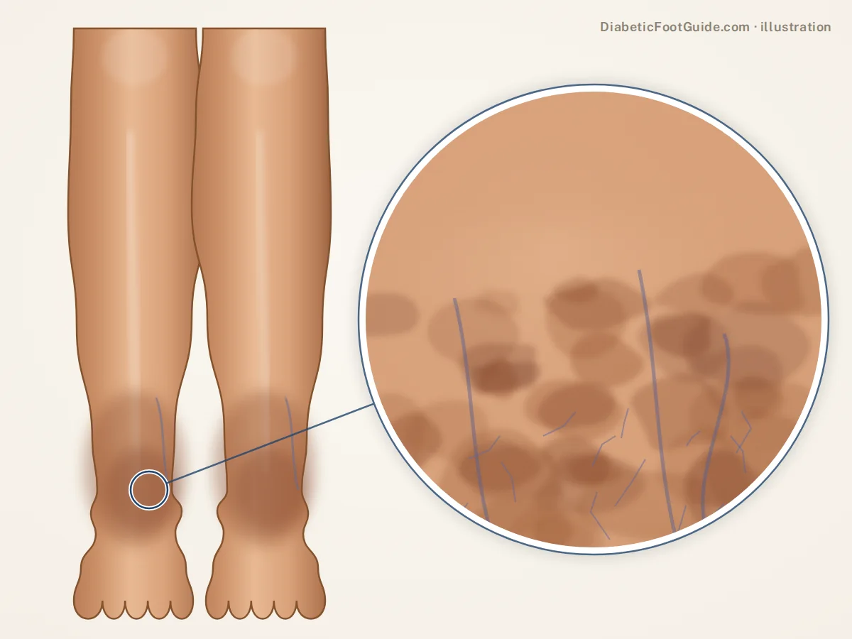
Venous ulcers announce themselves. The skin changes for months before it breaks, while this is still preventable.
-
Swelling that comes and goes
A bigger ankle by evening, better by morning, sock marks, nothing painful. Common on its own, but the pressure behind it drives what follows.
-
Brown staining and an itch
Iron leaks from overloaded veins and stains the skin rust-brown, which turns dry, scaly and itchy. This stasis eczema gets treated as athlete’s foot for a year, and scratching it is how many ulcers start.
-
Skin that turns hard and tight
A firm, shiny, tender band above the ankle, sometimes with small white scars. The ankle narrows while the calf stays full — an upside-down bottle. This skin tears easily.
-
The break
Usually a knock, a scratch or a burst blister. Instead of scabbing and shrinking it stays shallow and wet, with an irregular edge and a pink or yellow base. Two weeks on, the same size.
Arterial ulcers give less warning. The leg looks starved rather than congested: cool, shiny, hairless skin, thick nails, a foot that pales when raised and goes dusky red hanging down. The first sign is often a small dry sore that never granulates. Pain worse in bed and better hanging down is classic, though neuropathy erases it.
Not every mark on a diabetic shin is an ulcer
Round brownish sunken patches are usually diabetic dermopathy, which does not break down. Shiny yellow-brown plaques with visible small vessels are necrobiosis lipoidica, which can. Swelling with no wound is the swollen feet and legs stage, and the easiest to act on. Our picture guide shows them.
Venous, arterial or neuropathic: telling them apart
| Where it sits | What it looks like | Pain | Skin around it | What treats it | |
|---|---|---|---|---|---|
| Venous — most leg ulcers | Gaiter area, above the inner ankle | Shallow, wet, irregular edge | Aching, better with the leg raised | Swelling, rust-brown staining, eczema, varicose veins | Compression, once the arteries are checked |
| Arterial | Shin, outer ankle, heel, toes — where blood arrives last | Small, punched out, dry, pale base | Often severe. Worse raised, better hanging down — unless neuropathy masks it | Cool, shiny, hairless; weak or absent pulses | Restoring blood flow, not compression |
| Neuropathic — a foot ulcer, not a leg ulcer | Sole, ball of the foot, toe tips | Round, punched out, ringed with callus | Usually none, which is why it is found late | Callus, dry skin, deformity; good pulses | Pressure off — see diabetic foot ulcers |
Mixed ulcers are common, which makes this a measurement rather than a guess: valves and arteries fail together, and the wound looks venous while the circulation behaves arterial. Pressure ulcers and rarer causes are in ulcers that are not diabetic.
Diabetes complicates this twice. Neuropathy strips the pain out of an arterial ulcer, so “it does not hurt” rules nothing out; and roughly half of people with diabetes and a foot ulcer have peripheral artery disease.
Other sores on a diabetic leg
A knock on the shin that never healed
Skin over the shin is thin and short of spare circulation. On an older diabetic leg a minor wound there can simply stop and turn chronic. Two weeks without progress is the point to have it seen.
Necrobiosis lipoidica
Shiny yellow-brown plaques with a thinned center and visible small vessels, nearly always on the front of the lower legs and linked to diabetes. They ulcerate readily after minor injury.
The uncommon ones that change the plan
Pressure injury where a heel, cast edge or bed rail presses. Vasculitis. Pyoderma gangrenosum, which debridement worsens. And skin cancer, which can grow in a long-standing wound or present as one.
One rule covers them all: an ulcer that has not measurably improved after four to six weeks of correct treatment needs its diagnosis questioned rather than its dressing changed, and a wound with a rolled edge or tissue that bleeds easily needs a biopsy.
Should a leg ulcer be wrapped or compressed?
If it is venous, yes, and properly. Compression is not an accessory to the dressing, it is the treatment: guidelines put an active venous ulcer into multilayer or inelastic compression at around 40 mmHg at the ankle.
The same guidelines require arterial assessment first, and advise against sustained compression when ankle pressure is below 60 mmHg, toe pressure below 30 mmHg, or the index below 0.6. Compressing a leg already short of blood turns a slow wound into a dying one.
Why circulation is measured twice in a diabetic leg
Long-standing diabetes calcifies artery walls, so the cuff cannot close them and the index reads normal on a leg with poor flow. Toe arteries calcify far less, which is why toe pressures are used alongside it. Everyday hosiery is a different product — see diabetic socks versus compression socks.
What actually heals diabetic leg ulcers
- Name the cause first. Compression for venous, restored flow for arterial, pressure relief for neuropathic. A dressing chosen before that is a guess.
- Compression applied by someone trained, and kept on — including after the ulcer closes, which is what prevents the next one.
- Revascularization where inflow is the problem. Below a certain perfusion nothing heals, and no dressing changes that.
- Debridement and plain moist wound care. No dressing heals an ulcer by itself; advanced options are for wounds that stall despite good care. See healing a wound faster.
- Infection judged on what the leg is doing — spreading redness, warmth, new pain, pus, odor — not on a swab. See diabetic foot infection.
- The parts you control. Legs above heart level several times a day, ankle pumps and walking to work the calf pump, glucose down, and no smoking — nothing damages an arterial leg faster.
- Treat the veins, not just the wound. In a trial of 450 people, treating the faulty veins early alongside compression healed ulcers in a median of 56 days against 82, and ulcers later recurred at a lower rate.
Expect months, not weeks: in that trial half the compression-first group healed by twelve weeks, three-quarters by twenty-four. An arterial ulcer’s clock starts on the day blood flow improves.
Be seen promptly if
- Emergency: a cold, pale, mottled or dusky leg, or severe pain at rest — blood is not arriving
- Emergency: black tissue at the edge, crackling under the skin, or redness climbing the leg with fever
- Emergency: heavy bleeding from a vein — lie down, raise the leg above the heart and press firmly
- Same day: pus, a foul smell, a wound that has suddenly enlarged, or unmanageable blood sugars
- Same day: new one-sided calf swelling and tenderness, possibly a clot
- Within days: any new break below the knee that has not started to close
Common questions
What does an early diabetic leg ulcer look like?
Before it opens, the skin above the inner ankle changes: evening swelling, a rust-brown stain, dry itchy patches, then a firm shiny area. The ulcer itself starts shallow after a knock, with an irregular edge and a wet base that will not scab over.
Are leg ulcers caused by diabetes?
Usually not directly. Most are caused by vein disease or narrowed arteries, both commoner in diabetes; diabetes then slows healing, blunts the warning pain and raises infection risk. The neuropathic ulcer that people call diabetic forms on the foot, not the leg.
Why won’t a sore on my leg heal?
Four reasons cover most of it: arterial supply never measured, venous pressure never controlled with real compression, infection in the wound or the bone, or the wrong diagnosis. Wounds that resist correct treatment occasionally prove to be something else.
Should a leg ulcer be wrapped or compressed?
A venous one should be, and it is the most effective treatment there is — but not before the arteries are measured. Guidelines advise against sustained compression when ankle pressure is under 60 mmHg, toe pressure under 30 mmHg, or the index under 0.6.
How long do leg ulcers take to heal?
Months. In trial conditions about half of venous ulcers close by twelve weeks and three-quarters by twenty-four, with proper compression. Arterial ulcers depend on restoring blood flow. Anything that has not shrunk in four to six weeks needs reassessment, not a new dressing.
When is a leg sore an emergency?
A cold, pale, mottled or dusky leg, or severe pain at rest, means blood is not arriving: an emergency. So is spreading redness with fever, black tissue at the edge, or heavy bleeding from a vein. New one-sided calf swelling needs same-day assessment.
Sources
Clinical literature retrieved from PubMed, plus the governing guidance.
- Gohel MS, Heatley F, Liu X, et al. A Randomized Trial of Early Endovenous Ablation in Venous Ulceration. N Engl J Med. 2018;378(22):2105-2114. doi:10.1056/NEJMoa1801214 — median healing of 56 days with early vein ablation against 82 deferred, and 24-week healing of 85.6% against 76.3%, in legs fit for compression.
- Gohel MS, Mora J, Szigeti M, et al. Long-term Clinical and Cost-effectiveness of Early Endovenous Ablation in Venous Ulceration. JAMA Surg. 2020;155(12):1113-1121. doi:10.1001/jamasurg.2020.3845 — recurrence at 0.11 against 0.16 per person-year after early ablation, with no clear difference in time to a first recurrence.
- De Maeseneer MG, Kakkos SK, Aherne T, et al. ESVS 2022 Clinical Practice Guidelines on the Management of Chronic Venous Disease of the Lower Limbs. Eur J Vasc Endovasc Surg. 2022;63(2):184-267. doi:10.1016/j.ejvs.2021.12.024 — arterial assessment before compression, about 40 mmHg at the ankle, the thresholds below which compression is withheld.
- Fitridge R, Chuter V, Mills J, et al. The intersocietal IWGDF, ESVS, SVS guidelines on peripheral artery disease in people with diabetes and a foot ulcer. Diabetes Metab Res Rev. 2024;40(3):e3686. doi:10.1002/dmrr.3686 — roughly half of people with diabetes and a foot ulcer have peripheral artery disease, and how perfusion is judged when ankle pressures mislead.
- Mekkes JR, Loots MA, Van Der Wal AC, et al. Causes, investigation and treatment of leg ulceration. Br J Dermatol. 2003;148(3):388-401. doi:10.1046/j.1365-2133.2003.05222.x — the main causes of leg ulceration, and the less frequent ones including vasculitis, malignancy and pyoderma gangrenosum.
- Reid SD, Ladizinski B, Lee K, et al. Update on necrobiosis lipoidica: a review of etiology, diagnosis, and treatment options. J Am Acad Dermatol. 2013;69(5):783-791. doi:10.1016/j.jaad.2013.05.034 — plaques on the front of the lower legs, ulceration as a common complication, and watching for squamous cell carcinoma.
Related guides
Ulcers that are not diabetic
The same families, on the foot.
Healing a diabetic wound faster
What speeds a wound up, and what does not.
Diabetic dermopathy
The brown shin patches mistaken for ulcers.
Swollen feet and legs
The stage before a venous ulcer.
Diabetic socks vs compression socks
Why the wrong one matters here.
A leg sore that won’t heal, in Michigan?
Balance Foot & Ankle assesses lower-leg and foot wounds in Howell and Bloomfield Hills, starting with a circulation check before any compression.

Dr. Tom Biernacki is a double board-certified podiatric foot and ankle surgeon practising in Michigan. He treats diabetic wounds, Charcot neuroarthropathy and at-risk feet daily, and writes here to put the clinical reasoning behind that care in front of the people living with it.